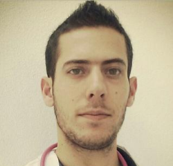
Mr Vitor Morgado-Weaver, Chief Cardiac Physiologist at Royal Devon and Exeter NHS Foundation Trust, reviews the recent BSE + ICE 2021 virtual meeting.
The challenges that came along with the COVID-19 pandemic have prompted us to think outside the box and re-invent the way we live, socialise, work and learn.
The British Society of Echocardiography has done an incredible job in supporting their members from the very early stages of the pandemic, working tirelessly to offer guidance for a safe but still high standard practice in echocardiography.
This year’s BSE + ICE meeting was different in the way it was delivered, but a great success from a learning point of view – the programme was diverse, rich and full of expert speakers in their areas who despite some technical issues, managed to deliver great informative talks. The chance to interact with sponsors was also a positive point of the meeting.
On day 1, I thoroughly enjoyed the session on aortic valve assessment by echocardiography. It is probably the valvulopathy that we most frequently come across in our clinics and because of its complexity with a multitude of considerations to take into account, the assessment is not always straightforward. As echocardiographers, we love measurements and parameters and aortic pathology is definitely one that requires us to use all the tools available to achieve the best diagnostic information for our patients. This should include 3D assessment, which could provide us with more accurate valve areas, and LV GLS with its role in risk stratification, and intervention guidance.
The second panel on cardiomyopathies was equally impressive and very comprehensive. With so many different types of cardiomyopathies, it is important to recognise relevant findings or signs that may suggest one in favour of another, and also use all the tools available for an adequate assessment.
Finally, to close the first day of the meeting we heard about interesting and relevant topics and novel applications that we should apply to our practice with a view of improving our patient care:
- GLS in cardio-oncology is a great technique with an important role in subclinical dysfunction and oncologic therapy guidance;
- Often it’s challenging to differentiate between an athlete’s heart and perhaps a mild phenotype of a cardiomyopathy and so we should assess these patients thoroughly, integrating the echo data and other imaging modalities;
- Novel applications of known techniques are of rising interest, including assessment of left atrium and right ventricle using strain analysis. Unfortunately mostly used in research at the present time and not widely available in all echo labs, which hopefully will change in the coming years!
Moving onto day 2 of the meeting, I have to say that even though I’m mostly an adult sonographer, I absolutely enjoyed the first panel on congenital heart disease. From a comprehensive overview on how to approach congenital patients, to assessment of AVSDs and causes of left heart obstruction. It’s so important to have a systematic approach when we come across these patients, even if not very often on our usual adult clinics. On a separate note, I shall not say functionally bicuspid AV ever again!
Session 2 on mitral valve assessment was quite interesting as well. Mitral stenosis is probably one of the valve lesions that we less frequently see, however with an aging population I suspect we will start seeing more calcific degenerative mitral stenosis and so it is important that we are comfortable assessing these patients.
Mitral regurgitation is a more commonly seen pathology in our clinics and often the TTE assessment alone will provide lots of diagnostic information, not only by using the standard 2D and hemodynamic assessment with Doppler, but also going further and explore the advantages of 3D echo. The mitral valve is a complex structure and often TTE’s have limitations for a number of reasons. This is where TOE becomes helpful for a comprehensive and detailed assessment of valve anatomy, severity of regurgitation and the mechanisms behind it. No mitral valve assessment should be complete without a 3D assessment of the valve – this provides an overview of the whole valve at once, and often we may be able to identify the problem straightaway. Image optimisation is key in any echocardiographic assessment and so your 3D will only be as good as your 2D images are, so always take time in assuring you optimise your images as much as possible. As every other technique, practice makes perfect!
To end up the last day of the meeting, we had some great cases in stress echo, both DSE and exercise and their advantages/usefulness in the contexts of ischaemia, MR and HCM. Sometimes patients report symptoms in exercise or clinicians believe there is a disparity between symptoms and echo findings, and that is where stress echo becomes a helpful diagnostic tool.
I often think of our job as playing detective – we look for clues or pieces of a bigger puzzle, analyse them and will hopefully be able to put them together and make sense of it. The diagnostic information we provide will only be as good as the knowledge we have and so, events like this are extremely important to facilitate continuous learning and development.
I hope that everyone enjoyed this year’s meeting as much as I did. I’d like to say a big thank you to all the speakers, and to the British Society of Echocardiography and the Irish Cardiac Echo and Imaging Group for organising such a great event.
Interested in other virtual events?
View the event list